by Terrence Doherty, MD
“How often should I have my eyes examined?” This is one of the most common questions that I hear from my patients. The answer, of course, depends on many factors. These include age, medical history, family history, and the presence of any ocular disease. Certainly anyone who has an established eye disease or a family history of diseases should have a regular eye exam to ensure timely intervention if needed. The same holds true for those with certain medical problems such as autoimmune diseases or diabetes, since these diseases can greatly impact our ocular health. But what about those folks with “healthy” eyes who are experiencing no problems with their vision? If we are just talking about checking glasses in younger patients, then every 1-2 years until our mid twenties is probably a good basic guideline. Then as needed for changes in vision or worn out spectacles. But the aging eye is quite different.
As we get into our 40’s, 50’s and 60’s, we all begin to experience changes to our vision. This usually starts with changes to our natural lens inside the eye. With every passing year, the lens becomes denser and stiffer as its cells become more and more compact. The stiffness reduces the eye’s ability to focus up close, manifesting as a loss of our near vision and the need for bifocals or reading glasses. Years later, as our lens becomes even denser, light entering our eye becomes altered and blocked, leading to the common symptoms of decreased nighttime vision, glare, and trouble driving. At this point, the lens is labeled a “cataract.” When cataract symptoms become significant enough, the patient is often advised to undergo cataract surgery. This procedure is the most commonly performed surgery in the United States and in almost all cases it greatly restores vision and stabilizes the glasses prescriptions for many years. And it never has to be repeated! In many cases, the surgery will even eliminate the need for glasses for many activities. It is likely that many folks who are reading this have already gone through this experience.
But there is certainly more to the story. As the eye ages, it can develop more than just a cataract. Certain other ocular diseases become more likely as well and can develop even before a cataract. One of these diseases is glaucoma.
Glaucoma is a term that most of us have probably heard. Most people are even aware that it has something to do with pressure inside the eye. Some of us may know of family members who have this diagnosis and take eye drops, but are not sure how it impacts them or its level of severity. Still others may have no idea what it is or may get it confused with other things like cataracts.
Glaucoma is a form of optic nerve disease that is caused by, or strongly correlated to, increased pressure inside the eye. This nerve damage can lead to severe vision loss and even permanent blindness. The optic nerve stems from the back of the eye and connects to the brain. It is made up of approximately 1 million nerve fibers from the retina that are susceptible to damage from increased pressure inside the eye. When this occurs, a diagnosis of glaucoma is made. To treat and prevent damage, the eye pressure has to be lowered.
So what causes this increased pressure? Although sometimes it can be caused by trauma or other ocular problems, most forms of glaucoma have no underlying cause. This is called primary glaucoma. We all have a measureable pressure inside our eyes that allows it to keep its round shape. Measuring this pressure is very easy and is part of a normal eye exam. A normal eye pressure would be considered anything below 21 mmHg (millimeters of mercury).
When eye pressure measures above 21, this is considered ocular hypertension and is a risk factor for developing glaucoma. Some eyes can measure pressures in the 30’s, 40’s or even higher. And the higher the pressure, the higher the risk. Remember, however, that glaucoma is only presents if there is measureable nerve damage. So high eye pressure alone does not necessarily mean glaucoma.
Nerve damaged is detected in several ways. One way is by using sophisticated imaging of the nerve in the doctor’s office. The most common of these is a test called Ocular Coherence Tomography, or OCT. This imaging is usually very quick and simple for the patient and yields a great deal of useful information that can be monitored and followed over time. (show image of OCT) That means that it is not only useful for making a diagnosing glaucoma, it is useful for following progression during the treatment of glaucoma. With the OCT, the optic nerve, which looks like a round disc in the back of the eye, is divided into segments and the thickness of the nerve fibers is precisely measured. To help aid the analysis, healthy segments show up as green, while borderline and high-risk segments show up as yellow and red respectively. The thinner the nerve fibers, the more damage that has occurred from glaucoma.
The other most common (and most important) way of diagnosing and monitoring glaucoma is by testing the patients’ peripheral vision thorough a special test called a Visual Field. That is because the peripheral vision, not the central vision, is what usually begins to diminish first as the optic nerve becomes damaged by pressure. As glaucoma progresses, the peripheral vision becomes smaller and smaller, creating tunnel vision for the patient, which, in severe cases, can eventually involve the central vision. This test usually takes about 5-10 minutes per eye. The patient is positioned in front of a large concave screen and dim lights are presented in various areas of the peripheral vision. When the light is detected, the patient clicks a button. The patient is monitored to make sure that they continue to stare straight ahead and click the button only when the lights are presented. As with the OCT, this test is not only used for diagnostic purposes, but for monitoring progression.
Now here’s the scariest part about glaucoma: the damage that is done to the nerve CANNOT be reversed. It is permanent. What is also troubling is that glaucoma does not have a lot of symptoms. The eye pressure is not painful and the peripheral vision loss in the early stages can develop so gradually that it often goes unnoticed, especially since the other eye can make up the difference. This is why screening tests are so important. It is unfortunate how many times I have seen patients present to the office with little to no symptoms, but have advanced nerve damage and significant vision loss from glaucoma.
The American Academy of Ophthalmology recommends that every person between the ages of 55 to 64 get a routine eye exam every 1-2 years even if there are no problems with vision. Over 65, they recommend every 6-12 months depending on the health of the eye. For patients with a family history of glaucoma, this exam should begin at age 40 and should be monitored regularly over a lifetime. This is especially true for African Americans, who have a 7-8 x higher risk of developing glaucoma.
The good news is that eye doctors now have more tools to lower the eye pressure and treat glaucoma. The most traditional and most common way is with the use of prescription eye drops. These can work very well and they remain a very important option for doctors and patients. However, often times there is the need for more than one prescription to control the pressure adequately. In addition, many drops have to be taken multiple times throughout the day. They also can cause some burning or irritation and cost quite a bit of money if they are not well covered by insurance. Because of this there has been an increasing movement by many glaucoma specialist to focus more on the surgical treatment of glaucoma. This is especially true due to the development of MIGS or Minimally Invasive Glaucoma Surgery.
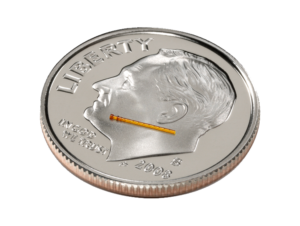
MIGS surgeries include a group of intraocular procedures that are indicated for mild to moderate glaucoma and are often done in conjunction with cataract surgery. However, some can also be done as primary procedures. The function of these operations is to improve and increase the drainage of fluid from inside the eye, which in effect will lower the intraocular pressure. Traditional glaucoma surgeries require making a fairly large incision into the wall of the eye, and although they are very effective in reducing the intraocular pressure, they have a long list of complications, including major infections. MIGS surgeries on the other hand, utilize microscopic sized drainage devices and very tiny incisions. And they are performed inside the eye. Thus, as the name implies, they are much less invasive and better maintain the eye’s integrity. MIGS surgeries are also relatively quick and easy to perform by an experienced specialist. They have the major advantage of decreasing the risks of traditional glaucoma surgeries such as infection and discomfort and they have been shown to work very well. Most people who have MIGS performed show a substantial decrease in their eye pressure and can reduce and sometimes eliminate their need for eye drops.
MIGS operations can be divided into several categories: Trabecular Bypass, Micrtrabeculectomies, and Suprachoroidal shunts.
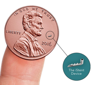
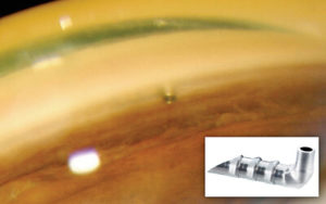
Trabecular Bypass surgeries work by bypassing the trabecular meshwork. This is the structure inside the eye’s natural drainage system that provides most of the resistance to outflow. It is located in the angle of the eye where the iris (the colored structure containing the pupil) meets the sclera (the white wall of the eye). The Kahook Dual Blade by New World Medical excises a large portion of the trabecular meshwork through a very small incision. This exposes the underlying collector channels that carry the fluid away through the blood vessels. The iStent from Glaukos is the other major form of bypass surgery. It is a tiny snorkel like device that is inserted through the trabecular meshwork to bypass it and gain direct access to the collector channels, thereby allowing better drainage.
Microtrabeculectomies utilize the same concept as traditional glaucoma surgeries (trabeculectomies) to form channels connecting the inside of the eye to a space underneath the conjunctiva, the transparent skin of the eye. The major difference being that the channels and the surgery are, once again, created from an internal approach, leaving the eye more intact and less prone to complications. The Xen Gel Stent is a small flexible tube that is inserted from the inside of the eye to this space under the conjunctiva, allowing the surgery to be faster and safer than a traditional trabeculectomy. It was recently approved by the FDA near the end of 2016 and has shown great promise in its early reports.
Suprachoroidal shunts include a device called the Cypass Micro Stent from Alcon. Like the Xel Gel stent, it is a small tube like device that inserts from the inside of the eye. However, it is designed to pass into a deeper area of the eye called the suprachoroidal space. This area is just underneath the scleral wall of the eye and contains a very large number of vascular capillaries. When the Cypass is inserted, it allows the fluid in the eye to flow into this rich vascular area where it enters the blood stream and is circulated away from the eye, lowering the pressure. Like the iStent, the Cypass is only approved by the FDA to be done in conjunction with cataract surgery at this time.
Certainly, MIGS will not completely replace the need for drops or traditional glaucoma surgeries. In some cases, these will still remain the best option. But MIGS does indeed fill a major gap in our treatment paradigm for glaucoma, namely, treating those that require some form of treatment or medication for glaucoma, but are below the threshold for needing more aggressive surgical treatment. Perhaps the best part is that most of the time MIGS surgeries are done at the same time as cataract surgery, so there are no additional trips to the operating room to treat both. This is both cost saving and convenient.
So for those of you out there who have a diagnosis of glaucoma and require medication, consider talking to your doctor about the newer treatments for lowering eye pressure such as MIGS, especially if you have never had cataract surgery. And if you have never had a conversation with your eye doctor about glaucoma, you should consider making an appointment in the near future to measure your level of risk. It could be one of the most important decisions you could ever make.
